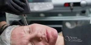
Facial skin diagnostic
Analysis of skin condition allows you to analyze the depth and prevalence of pathologies such as dilated blood vessels, latent pigmentation, acne porphyrins, pores, texture, keratinization, wrinkles, and correct prognosis of results and draw up an optimal treatment plan.

Modern diagnostic system OBSERV 520 in our clinic allows us to assess pigmentary pathology, vascular pathology, microrelief, wrinkles, pores, texture, skin uniformity, as well as to reveal latent pigmentation and blood vessels.
Pigmentation
Pigment spots are a collective term in aesthetic medicine, which includes a large number of pathologies of pigment metabolism disorders in the direction of increasing the production of melanin - the pigment that determines the color of the skin. Pigmented spots include solar lentigo, ephelids (freckles), melasma, chloasma, post-inflammatory hyperpigmentation and other skin pathologies.
Each problem has its own causes and depth of pigment. Accordingly, treatment approaches will be different. In some cases it will be photorejuvenation and/or fractional laser resurfacing, and/or Jessner peels professional cosmetics. Analysis of the condition of the skin will allow you to accurately determine the tactics and predictions of treatment. The system uses ultraviolet rays of a specific wavelength, which is well absorbed by melanin. Clusters of melanin will appear darker and more contrasting than normal daylight. This makes it possible to detect even the smallest changes in the concentration of pigment in the skin and to diagnose pigmentation disorders in the early stages. Timely diagnostics will help fix the problem at an early stage.
Hidden pigmentation
Latent pigmentation also becomes more visible in ultraviolet rays. This allows in the early stages to diagnose the increase in the formation of melanin (pigment) even before the appearance of age spots on the surface. Like the hidden, subtle pigmentation on the cheek in the photo above. The combination of laser treatments and the right home care will help control and prevent overt pigmentation.
Acne in ultraviolet rays
Patented ultraviolet light analysis of acne detects the acne pathogen, Propionibacterium Acnes. Depending on the concentration of bacteria, a glow of different colors appears. This allows you to determine the patency of pores, identify areas of maximum damage and choose tactics acne treatment or facial skin care.

Subsequent photos will allow you to evaluate in dynamics the correctness of the chosen treatment tactics, and, if necessary, adjust the treatment.
Vascular pathology
Like age spots, the aesthetic problem of dilated vessels of the skin of the face - rosacea (telangiectasia) has its own characteristics depending on the anatomical zone and depth of occurrence, the presence of collaterals. One of the modes is the analysis of vascular pathology. Thanks to this, it is possible to see even those vessels that are not visible to the naked eye during a normal examination of the skin. This will allow the doctor to choose the scope of the procedure and the optimal method: laser vascular removal, photocoagulation or sclerotherapy. Coagulation of vessels in the early stages allows you to achieve lasting results. In patients prone to rosacea, this method can identify areas with labile capillaries.
Relief and texture
The state of the surface layer of the skin determines the microrelief - irregularities on the skin. Disturbance of microrelief can be caused by various factors - age, lifestyle, skin type, improperly selected cosmetics and other factors. A special analysis mode will show all texture irregularities and the most problematic areas of the skin that require increased attention.
- Enlarged pores
- Disorder of skin keratinization
- Wrinkles
Depending on this, the direction of treatment can vary from a banal selection of cosmetics to treatment with professional means and treatment of concomitant diseases. You can track the dynamics of microrelief changes from the very beginning of the treatment.
Fungal lesions
A special study mode - Wood's lamp, has proven itself well in dermatology. Wood's lamp is also called a black light lamp, since this spectrum of ultraviolet radiation is not visible to our eyes. Under the influence of ultraviolet rays of this spectrum, fluorescence of various biological molecules of a substance is observed. This is a standard analysis method that has been used in dermatology for many years and allows you to detect dermatophytosis and trichophytosis - fungal lesions of the skin and hair, to diagnose the depth of pigmentation.
Dyschromia
Dyschromia is an irregularity in the color of the skin, which our eye distinguishes as an uneven color. Dyschromia, on closer inspection, is an alternation of areas of hyperemia (redness) and pigmentation, which creates for our eyes a feeling of a heterogeneous skin tone. A special study mode will allow you to identify these problem areas and select the optimal treatment plan. After the course of procedures, you can assess the condition and uniformity of skin tone.
Photos before and after procedures
Evaluation of the effectiveness of treatment in dermatology and cosmetology is the cornerstone. Skin changes occur rather slowly over several weeks, or even months. All this happens in front of the patient's eyes and, looking at ourselves every day in the mirror, we sometimes do not notice dramatic changes, since this happens slowly and in our eyes. Therefore, it can be difficult to trace the difference before and after the procedure. One of the advantages of analyzing the skin of the face in our clinic is that you can compare the photos before and after the treatment.
Price
Prices for skin analyzing in Kiev
| Price | Duration | |
| Doctor's consultation with Observ skin analysis | UAH 500 | 30 minutes |
| Follow-up consultation | 250 UAH | 20 minutes |
Frequently Asked Questions
Is it dangerous?
No, it is safe for the skin, eyes and the body as a whole. For the purpose of analysis, various modes of polarization of visible light are used. During the study, the intensity of ultraviolet radiation is several times less than the intensity of ultraviolet rays in the composition of solar radiation of daylight, which we encounter even on a cloudy day. Therefore, you should not be afraid of this research method, as well as repeated research - it is absolutely safe.
Is a doctor's consultation included in the price?
Yes, the cost of analyzing the skin of the face includes a doctor's consultation. The doctor will give you a detailed consultation on the condition of the skin and existing methods of correction.
Skin diagnostics is included in the cost of each procedure. If you do the procedure on the same day, then the cost of diagnostics and consultation with a doctor will be included in the cost of the procedure. During repeated visits for procedures, diagnostics will be included in the price and a separate fee will not be charged.
Is it possible to get the research result?
Yes, skin diagnostics involves sending a report by email. Please include your email address during your visit and the doctor will send you the results of the study.
How long are results stored?
Research results are stored for 1 year.
Is it possible to examine a mole?
No, this technique is aimed at studying the aesthetic parameters of the skin, such as pigmentation, the presence of porphyrins in acne, the presence and prevalence of dilated vessels (telangiectasias), fungal lesions, dyschromia and changes in the uniformity of skin tone.
Is it possible to detect Demodex using this method?
This technique does not allow to confirm qualitatively or quantitatively the presence of Demodex.


